
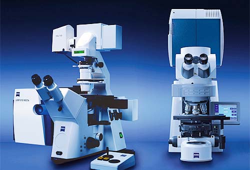
Zeiss LSM META
Manufacturer: Zeiss
Location: Britanya 233
Internal User Price: 35NIS/hour
External User Price: 350NIS/hour
Acquisition Date: 2006
Details
The unique scanning module is the core of the LSM 510 META. It contains motorized collimators, scanning mirrors, individually adjustable and positionable pinholes, and highly sensitive detectors including the META detector. All these components are arranged to ensure optimum specimen illumination and efficient collection of reflected or emitted light. A highly efficient optical grating provides an innovative way of separating the fluorescence emissions in the META detector. The grating projects the entire fluorescence spectrum onto the 32 channels of the META detector. Thus, the spectral signature is acquired for each pixel of the scanned image and subsequently can be used for the digital separation into component dyes.
Established: 2006
Configuration: inverted
XYstage: motorized, with universal holder or incubator chamber
Z-drive: motorized, minimal step 50 nm
Objectives: x10; x25 (universal immersion – water or glycerol or oil); x40 (water immersion); x63 (oil immersion); x100 (oil immersion) assembled on motorized revolver
Transmitted light condenser: manual with DIC
Lasers: Argon 458, 488 and 514 nm; output 30 mW, tunable;
DPSS 561 nm; output 15 mW;
HeNe2 633 nm; output 10 mW
Diode 405 nm; output 30 mW;.
Detectors: 2 PMTs each with adjustable pinhole and changeable filters, META detector with adjustable pinhole and one PMT for the transmitted light.
Special features: META detector – provides spectral sensitivity. Light from a sample dispersed by a grating reaches a line of 32 small PMTs.
Spectral resolution about 10 nm.
Autofocus.
Specialized imaging techniques: multiple ROI, bidirectional scanning, tile acquisition, z-stack, time series, photobleaching (FRAP), energy transfer (FRET), emission fingerprinting, spectral unmixing for fluorophores with close emissions or for effective background reduction, live cell imaging. 5-D acquisitions –X, Y, Z, time and lambda.
Additional equipment: Removable incubator chamber for cell maintenance and viewing with temperature, CO2 and humidity control.